
Understanding an ER visit
Imaging in the ER: CT, X-ray and Ultrasound Explained
Published September 12, 2026
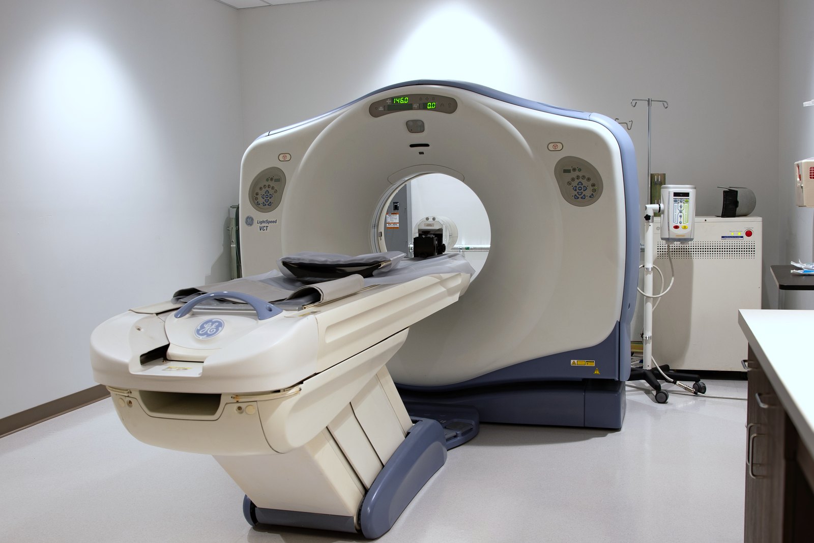
Having CT and X-ray in the building, and ultrasound a call away, is the practical difference between an emergency room and an urgent care — not because we are faster, but because the question you arrived with can be answered before you leave rather than referred onward.
This page explains what each one is for, what the radiation actually amounts to, and — the part most sites leave out — when a scan is not worth having.
CT and X-ray on site; ultrasound called in
The question you arrived with can be answered before you leave.
And when a scan is not worth it
The part most sites leave out, and it is on this page.
If you are unsure, come in
This list is not exhaustive and you are not expected to diagnose yourself.
What each one is good at
X-ray
Bones and lungs. Fractures, dislocations, pneumonia, a collapsed lung, swallowed objects. Quick, low radiation, and usually the first picture taken for an injury.
What it cannot do: soft tissue. A normal X-ray does not rule out a ligament tear, and it frequently does not rule out a small fracture in the first days — which is why a normal film sometimes comes with advice to come back.
CT
Detail, and bleeding. Head injuries and suspected bleeds, abdominal pain, chest problems, complex fractures, clots in the lungs, kidney stones. It builds a cross-sectional picture, so it sees things a flat X-ray cannot.
It uses meaningfully more radiation than an X-ray, which is why it is chosen rather than defaulted to.
Ultrasound
Soft tissue, fluid, and blood flow. No radiation at all. Gallbladder, kidneys, the womb in pregnancy, clots in a leg, fluid where it should not be, and guiding a needle accurately. At El Mirage ER it is called in from a mobile service rather than kept in the building.
It is the first choice in pregnancy and often in children, precisely because there is no radiation involved. Its limitation is that it does not see through bone or air well, so it is poor for lungs and for the brain.
Radiation, answered honestly
No numbers here, and that is deliberate. A millisievert figure means very little without a comparison, and the comparisons that circulate are usually misleading. What is actually useful is the shape of it:
Ultrasound involves no radiation. None.
An X-ray involves a small amount — the sort of exposure that is a routine part of medicine and is not something to weigh against an actual diagnostic need.
A CT involves meaningfully more than an X-ray. Not dangerous for one scan when it is needed, and genuinely worth avoiding when it is not.
The way to think about it is not “is radiation bad” but “does this scan change what happens next.” A CT that finds a bleed is worth far more than the exposure costs. A CT done because everyone feels better having a picture is worth less than nothing — it carries the exposure, and it tends to find incidental things that lead to more tests.
You are allowed to ask why a scan is being suggested, what it will change, and whether there is an alternative. That is a reasonable question and not an awkward one. If you are pregnant or might be, say so before any imaging.
When a scan is not worth having
This is the section that gets left off most hospital and ER pages.
Most back pain. Adult spines commonly show disc and joint changes in people with no pain at all, so finding one in someone who hurts does not establish cause. More on back pain
Most headaches. The pattern matters far more than the picture, and a scan of an ordinary headache reassures nobody for long. More on headaches
Minor injuries with no bone tenderness and normal weight-bearing. More on sprains and breaks
Repeating a scan that was already normal, when nothing has changed.
“We are not going to scan that” is sometimes the right answer, and it is not a brush-off. If it is said to you, asking what would make you change your mind is a good question — it gives you the thing to watch for at home.
Why having them here matters
An urgent care that suspects a fracture, a bleed, a clot or a stone has to send you to an emergency room, and the sequence starts again — new registration, new assessment, then the picture.
Here, the imaging and the laboratory are in the same building as the physician reading them, so the assessment, the picture and the decision happen in one visit. For some presentations that is a convenience. For a suspected bleed or a clot it is the whole point.
If you need a test we do not have here, or a specialist to interpret something, we arrange the transfer to a hospital. We are a freestanding emergency room, not a hospital, and knowing which questions we can close on site is more useful to you than a list of equipment.
What to tell us before imaging
If you are pregnant, or might be.
Any previous scans, especially recent ones of the same area.
Kidney problems, diabetes, or a previous reaction to contrast dye, since some CT scans use an injected dye.
Anything metal — implants, a pacemaker, shrapnel, surgical clips.
If you are worried about radiation. Say it. It is a reasonable thing to raise and it will change the conversation rather than end it.
Sources
The guidance in this article about when to seek emergency care is drawn from these public sources. Each one is linked so you can read it yourself.
MedlinePlus (National Library of Medicine) — CT Scans — medlineplus.gov/ctscans.html
This article is general information, not a diagnosis, and it cannot account for your history or medications. If you are unsure, call us on (480) 237-4922 or come in. If it might be an emergency, treat it as one and call 911.
A note on this article: This information is general health education and is not a substitute for a visit with a provider. If you have a concern about your health or your family's health, call us and we will help.